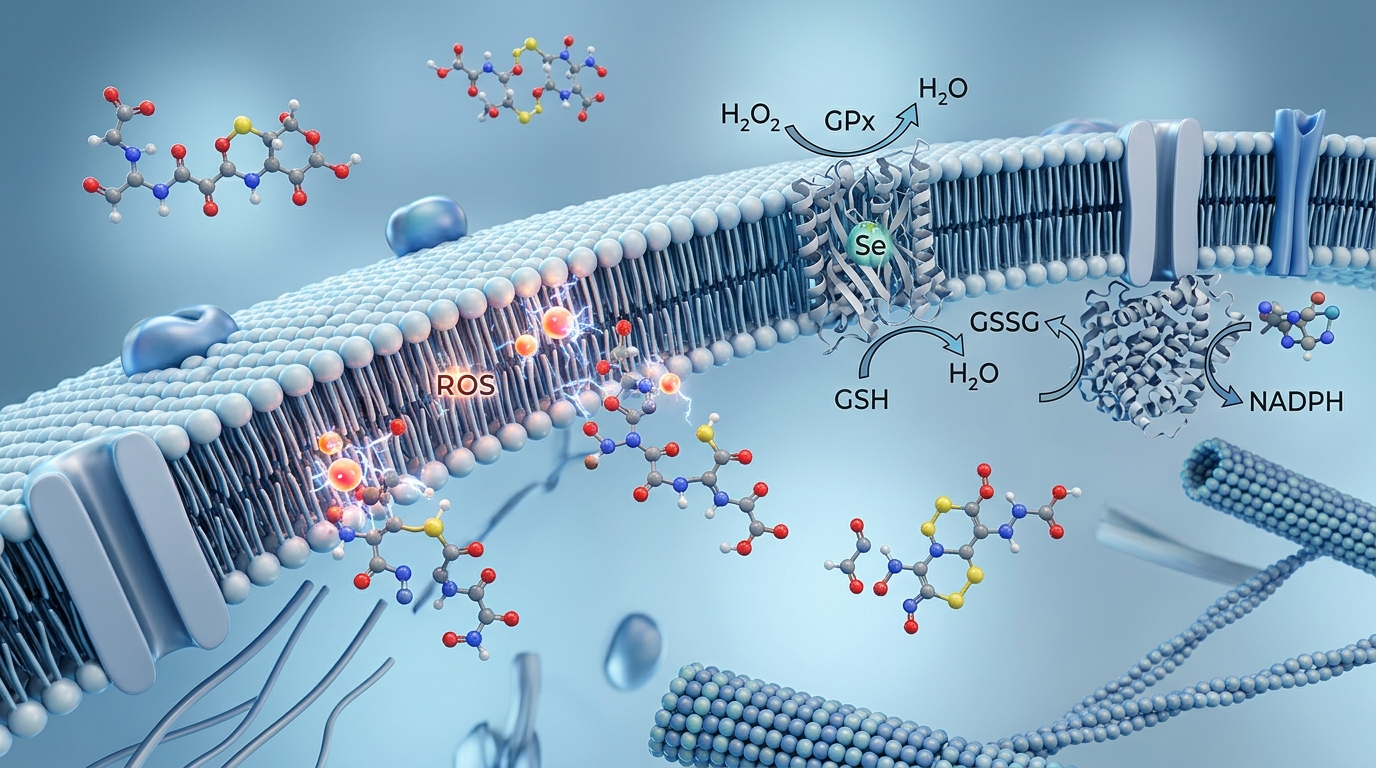
Main Conclusion: Glutathione functions as the foundational endogenous tripeptide regulator of cellular redox homeostasis, exerting critical biological control over oxidative stress defense, Phase II hepatic biotransformation, and immune-mitochondrial bioenergetics across experimental and clinical research models.
This overarching conclusion is established through three core scientific mechanisms:
- Direct Intracellular Redox Buffering and Enzymatic Defense: Glutathione neutralizes reactive oxygen species directly and acts as the indispensable cofactor for protective antioxidant enzymes.
- Substrate-Driven Phase II Biotransformation: Glutathione conjugates electrophilic xenobiotics and endogenous metabolites to facilitate cellular export and clearance.
- Regulation of Mitochondrial Bioenergetics and Immune Signaling: Glutathione maintains mitochondrial membrane integrity and modulates lymphocyte activation pathways.
1. Direct Intracellular Redox Buffering and Enzymatic Defense
Glutathione (γ-L-glutamyl-L-cysteinylglycine, or GSH) is the most abundant non-protein thiol in eukaryotic cells, maintained at millimolar concentrations (1–10 mM) to preserve a reducing intracellular environment.
In-Vitro Evidence
- Cell-free and cell culture assays demonstrate that GSH directly scavenges hydroxyl radicals, singlet oxygen, and lipid peroxides via hydrogen donation from its cysteine sulfhydryl (-SH) group, forming oxidized glutathione disulfide (GSSG).
- In-vitro kinetic analyses confirm GSH functions as an obligatory electron donor for Glutathione Peroxidase (GPx) enzymes in reducing hydrogen peroxide to water, maintaining a high GSH:GSSG ratio (>100:1 in healthy resting cells).
Animal Evidence
- Murine models with genetically knocked-out glutamate-cysteine ligase catalytic subunit (GCLC)—the rate-limiting enzyme in GSH synthesis—exhibit embryonic lethality due to widespread oxidative apoptosis and tissue degradation.
- Rodent models of chemically induced oxidative stress demonstrate that exogenous GSH replenishment restores endogenous GPx activity and attenuates lipid peroxidation markers (malondialdehyde).
Human Clinical Evidence
- Randomized controlled trials (RCTs) evaluating oral, sublingual, and liposomal GSH supplementation have shown statistically significant increases in total body glutathione stores and reductions in systemic oxidative stress biomarkers, such as plasma 8-isoprostane.
- Clinical observational studies consistently demonstrate that depressed plasma and erythrocyte GSH:GSSG ratios strongly correlate with advanced biological aging and chronic metabolic pathologies.
Hypotheses and Research Frontiers
- Researchers hypothesize that targeted delivery of synthetic GSH peptide analogs to specific cellular compartments could provide neuroprotective effects in neurodegenerative models characterized by localized redox collapse.
2. Substrate-Driven Phase II Biotransformation
Glutathione plays an essential role in hepatic biotransformation, converting reactive and lipophilic compounds into water-soluble conjugates for biliary and renal excretion.
In-Vitro Evidence
- Isolated hepatocyte assays show that Glutathione S-Transferases (GSTs) catalyze the nucleophilic attack of the GSH thiol group onto electrophilic carbon, nitrogen, or sulfur atoms of xenobiotics, neutralizing their mutagenic potential.
- In-vitro transport assays confirm that multidrug resistance-associated proteins (MRPs) actively transport GSH-electrophile conjugates out of cells across the plasma membrane.
Animal Evidence
- Toxicological studies in rodent models illustrate that acetaminophen (APAP) overdose rapidly depletes hepatic GSH pools, leading to toxic accumulation of N-acetyl-p-benzoquinone imine (NAPQI) and subsequent centrilobular necrosis; GSH precursor administration completely halts this cascade.
- In-vivo metabolic tracing in rats confirms the degradation of GSH-conjugates in the kidneys into mercapturic acids, which are efficiently eliminated via urine.
Human Clinical Evidence
- Clinical data from emergency medicine robustly establish that supporting GSH synthesis through N-acetylcysteine (NAC) prevents fatal hepatic necrosis following toxic electrophilic xenobiotic exposures.
- Genetic association studies indicate that human polymorphisms resulting in GST deletions (such as GSTM1-null phenotypes) impair Phase II clearance capacity and increase vulnerability to toxicological damage.
3. Regulation of Mitochondrial Bioenergetics and Immune Signaling
Glutathione is vital for safeguarding mitochondrial DNA and tuning the functional responsiveness of immune cells.
In-Vitro Evidence
- Isolated mitochondrial preparations reveal that mitochondrial GSH (mGSH), which accounts for 10–15% of total cellular GSH, is critical for preventing cytochrome c release and preserving inner mitochondrial membrane polarization.
- Cell culture studies of human T-lymphocytes show that intracellular GSH depletion impairs interleukin-2 (IL-2) receptor expression and prevents antigen-induced lymphocyte blastogenesis.
Animal Evidence
- Transgenic animal studies show that selective depletion of mGSH sensitizes hepatocytes to tumor necrosis factor-alpha (TNF-α)-mediated apoptosis, even when cytosolic GSH levels are partially preserved.
- Murine infectious disease models indicate that maintaining cellular GSH levels improves macrophage phagocytic capacity and dampens excessive nuclear factor kappa B (NF-κB) pro-inflammatory signaling.
Human Clinical Evidence
- Clinical trials of oral and liposomal GSH administration in healthy adults demonstrate a direct enhancement of Natural Killer (NK) cell cytotoxicity and a dose-dependent increase in lymphocyte proliferation.
- Patients with chronic viral infections and associated immune dysfunction frequently present with severe GSH deficiency; restoration of thiol pools has been clinically linked to improved immune markers.
Anecdotal and Unverified Claims
- Anecdotal Claim: Intravenous (IV) glutathione is widely promoted in aesthetic settings for rapid skin lightening and systemic detoxification.
- Scientific Context: While IV administration rapidly increases transient circulating thiol levels, robust, double-blind clinical evidence validating long-term safety, optimal therapeutic dosing, and durable aesthetic outcomes remains limited, prompting regulatory warnings regarding unapproved systemic administration.