Key Takeaways
- Structural Origin: KPV (Lys-Pro-Val) represents the carboxy-terminal tripeptide fragment (residues 11–13) of alpha-melanocyte-stimulating hormone (α-MSH).
- Mechanistic Divergence: While full-length α-MSH acts predominantly via G protein-coupled melanocortin receptors (MC1R–MC5R), KPV operates largely through receptor-independent, transporter-mediated intracellular pathways.
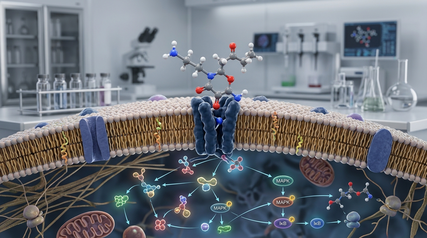
- Transport & Signaling: KPV is internalized via the oligopeptide transporter PepT1 (SLC15A1) and inhibits nuclear factor-kappa B (NF-κB) and MAP kinase activation at nanomolar concentrations.
- Absence of Pigmentary Effects: Because KPV does not activate MC1R melanogenic pathways, it avoids the pigmentary changes associated with α-MSH and synthetic melanocortin agonists.
- Translational Status: Despite robust preclinical evidence in models of intestinal, pulmonary, and cutaneous inflammation, KPV has not completed formal human clinical trials and is not approved by the FDA.
The Melanocortin System and the Genesis of KPV
The melanocortin system is a central neuroendocrine network comprising peptide ligands derived from the proopiomelanocortin (POMC) precursor, including adrenocorticotropic hormone (ACTH) and α-, β-, and γ-melanocyte-stimulating hormones (α-, β-, γ-MSH). These endogenous agonists exert their physiological actions through five distinct G protein-coupled receptors designated MC1R through MC5R. Beyond its canonical role in regulating pigmentation and melanogenesis through MC1R, α-MSH functions as a potent endogenous mediator of immune tolerance and anti-inflammatory counter-regulation.
Full-length α-MSH is a 13-amino-acid neuropeptide (Ac-Ser-Tyr-Ser-Met-Glu-His-Phe-Arg-Trp-Gly-Lys-Pro-Val-NH2). Therapeutic exploration of native α-MSH has long been constrained by two pharmacological hurdles: a short biological half-life due to rapid enzymatic degradation, and non-selective receptor activation leading to cutaneous hyperpigmentation and systemic effects. Structural-functional mapping demonstrated that the anti-inflammatory properties of α-MSH could be localized to its C-terminal sequence, leading to the isolation and characterization of the tripeptide Lys-Pro-Val, commonly known as KPV.
Mechanistic Divergence: Classical Melanocortin GPCRs vs. KPV
The defining scientific interest in KPV melanocortin signaling lies in how this tripeptide diverges mechanistically from full-length α-MSH and other synthetic melanocortin analogs. Classical melanocortin signaling depends on receptor binding at the plasma membrane, eliciting intracellular cyclic adenosine monophosphate (cAMP) accumulation through adenylate cyclase stimulation. By contrast, laboratory investigations reveal that KPV relies on fundamentally distinct cellular entry and signaling mechanisms.
1. Absence of MC1R-Driven Melanogenesis
The central pharmacophore for melanocortin receptor activation resides in the core His-Phe-Arg-Trp (residues 6–9) sequence of α-MSH. Because KPV lacks this binding sequence, it does not stimulate the MC1R-cAMP-tyrosinase cascade responsible for melanin synthesis. This absence of melanogenic activity permits the targeted investigation of anti-inflammatory cascades without confounding skin pigmentation responses.
2. Transporter-Mediated Internalization via PepT1
Rather than relying primarily on cell-surface GPCR engagement, KPV utilizes active peptide transport systems. Landmark work in intestinal epithelial models revealed that KPV is transported across cell membranes by the proton-coupled oligopeptide transporter 1 (PepT1, encoded by SLC15A1). PepT1 is expressed under baseline conditions in the small intestine and is markedly upregulated in the colonic epithelium during active inflammatory states, such as inflammatory bowel disease (IBD). This allows KPV to preferentially enter inflamed epithelial and immune cells.
3. Direct Intracellular Modulation of NF-κB and MAPK
Once internalized, KPV suppresses canonical pro-inflammatory signaling pathways. In vitro studies show that nanomolar concentrations of KPV inhibit the phosphorylation and degradation of IκB-alpha, preventing the nuclear translocation of the p65 subunit of nuclear factor-kappa B (NF-κB). Concurrently, KPV attenuates mitogen-activated protein kinase (MAPK) cascades, leading to down-regulation of pro-inflammatory cytokines including TNF-α, IL-1β, and IL-6. Studies in bronchial epithelial models indicate that C-terminal melanocortin fragments may interfere with nuclear transport proteins (such as importins), underscoring a mechanism of action centered inside the cell rather than on the cell surface.
Preclinical Evidence Across Experimental Models
Preclinical evaluation of KPV spans gastrointestinal, pulmonary, and dermatological disease models, establishing its biological profile across various organ systems.
Intestinal Inflammation and Mucosal Integrity
The most extensive body of evidence for KPV exists in rodent models of experimental colitis. In both dextran sulfate sodium (DSS)-induced and 2,4,6-trinitrobenzenesulfonic acid (TNBS)-induced colitis models, oral administration of KPV significantly reduced histological inflammation, decreased colonic myeloperoxidase (MPO) activity, and preserved epithelial architecture. Notably, studies utilizing mutant mice with non-functional MC1R receptors (MC1Re/e) demonstrated that KPV retained its therapeutic efficacy and rescued mice from colitis-induced mortality, confirming that its primary in vivo protective mechanism operates independently of MC1R signaling.
Airway and Cutaneous Inflammation
In human bronchial epithelial cell cultures, KPV significantly suppressed chemokine secretion and cytokine-driven inflammation. Similarly, in models of contact dermatitis and cutaneous vasculitis, topical and systemic application of α-MSH-related tripeptides diminished leukocyte infiltration, local edema, and tissue damage without inducing hyperpigmentation.
Antimicrobial Properties
In vitro investigations have identified antimicrobial actions for KPV against pathogens including Candida albicans and Staphylococcus aureus. Research indicates the tripeptide can disrupt membrane stability and modulate microbial proliferation, representing an auxiliary biological property distinct from host-directed immune pathways.
Research Limitations and Clinical Evidence Gaps
Despite promising in vitro and animal findings, significant scientific limitations remain regarding KPV:
- Lack of Controlled Human Data: The overwhelming majority of published research on KPV consists of preclinical cell culture and rodent studies. There are no completed, peer-reviewed, phase 2 or phase 3 randomized controlled trials evaluating KPV for any clinical indication.
- Pharmacokinetic Uncertainties: While PepT1 facilitates cellular uptake in intestinal tissue, systemic pharmacokinetics, oral bioavailability outside the gastrointestinal tract, and metabolic clearance profiles in humans have not been rigorously established in clinical settings.
- Potential Off-Target Effects: Although KPV does not activate MC1R melanogenesis, potential low-affinity interactions with other melanocortin subtypes or non-specific cellular targets require systematic mapping.
Regulatory and Developmental Status
KPV is an unapproved investigational research peptide. It has not been approved by the U.S. Food and Drug Administration (FDA), the European Medicines Agency (EMA), or any other national regulatory authority for the prevention, diagnosis, or treatment of any medical condition. It is available strictly for scientific and laboratory research purposes and should not be used in clinical practice or personal administration outside authorized investigative frameworks.
Frequently Asked Questions
How does KPV differ functionally from full-length α-MSH?
Full-length α-MSH activates all five melanocortin GPCRs (MC1R–MC5R), stimulating pigmentation via MC1R in addition to modulating immune pathways. KPV lacks the central melanocortin binding core; it does not induce melanogenesis and operates largely through PepT1-mediated cellular uptake and intracellular inhibition of NF-κB and MAPK.
What role does the PepT1 transporter play in KPV activity?
PepT1 (SLC15A1) is an oligopeptide transporter responsible for moving di- and tripeptides into cells. Because PepT1 expression increases during intestinal inflammation, KPV is transported efficiently into inflamed intestinal epithelial and immune cells, where it exerts targeted intracellular anti-inflammatory effects.
Is KPV FDA-approved for inflammatory bowel disease?
No. KPV is not FDA-approved for IBD or any other condition. Its protective effects have been documented in laboratory cell cultures and animal models of colitis, but it has not undergone formal clinical safety and efficacy evaluation in humans.
Does KPV cause skin tanning or pigment changes?
No. Preclinical studies show that KPV does not activate the MC1R-dependent melanogenic cascade responsible for skin darkening, which distinguishes it from parent α-MSH and synthetic melanocortin receptor agonists like Melanotan II.
References
- Dalmasso G, et al. PepT1-mediated tripeptide KPV uptake reduces intestinal inflammation. Gastroenterology. 2008;134(1):166-178.
- Brzoska T, et al. α-melanocyte-stimulating hormone and related tripeptides: biochemistry, antiinflammatory and protective effects in vitro and in vivo. Endocr Rev. 2008;29(5):581-602.
- Kannengiesser K, et al. Melanocortin-derived tripeptide KPV has anti-inflammatory potential in murine models of inflammatory bowel disease. Inflamm Bowel Dis. 2008;14(3):324-331.
- Land SC. Inhibition of cellular and systemic inflammation cues in human bronchial epithelial cells by melanocortin-related peptides: mechanism of KPV action and a role for MC3R agonists. Int J Physiol Pathophysiol Pharmacol. 2012;4(2):59-73.
- The Melanocortin System in Inflammatory Bowel Diseases: Insights into Its Mechanisms and Therapeutic Potentials. Int J Mol Sci. 2023;24(14):11603.
Research Summary
The research compound KPV exemplifies a distinct pharmacological divergence within the melanocortin family. By shedding the melanogenic pharmacophore of α-MSH, this tripeptide retains the capacity to downregulate NF-κB and MAPK inflammatory signaling via PepT1 transporter-mediated uptake rather than classical GPCR stimulation. Although animal models demonstrate substantial anti-inflammatory efficacy in gastrointestinal and epithelial models, the current evidence base remains entirely preclinical. KPV is not approved for human clinical use, and comprehensive clinical trials are required to determine its pharmacokinetic profile, safety parameters, and therapeutic potential in humans.