Key Takeaways
- Membrane permeability barrier: Intact NAD+ is a large, polar, phosphorylated dinucleotide that cannot readily cross mammalian plasma membranes without specialized transport or enzymatic cleavage.
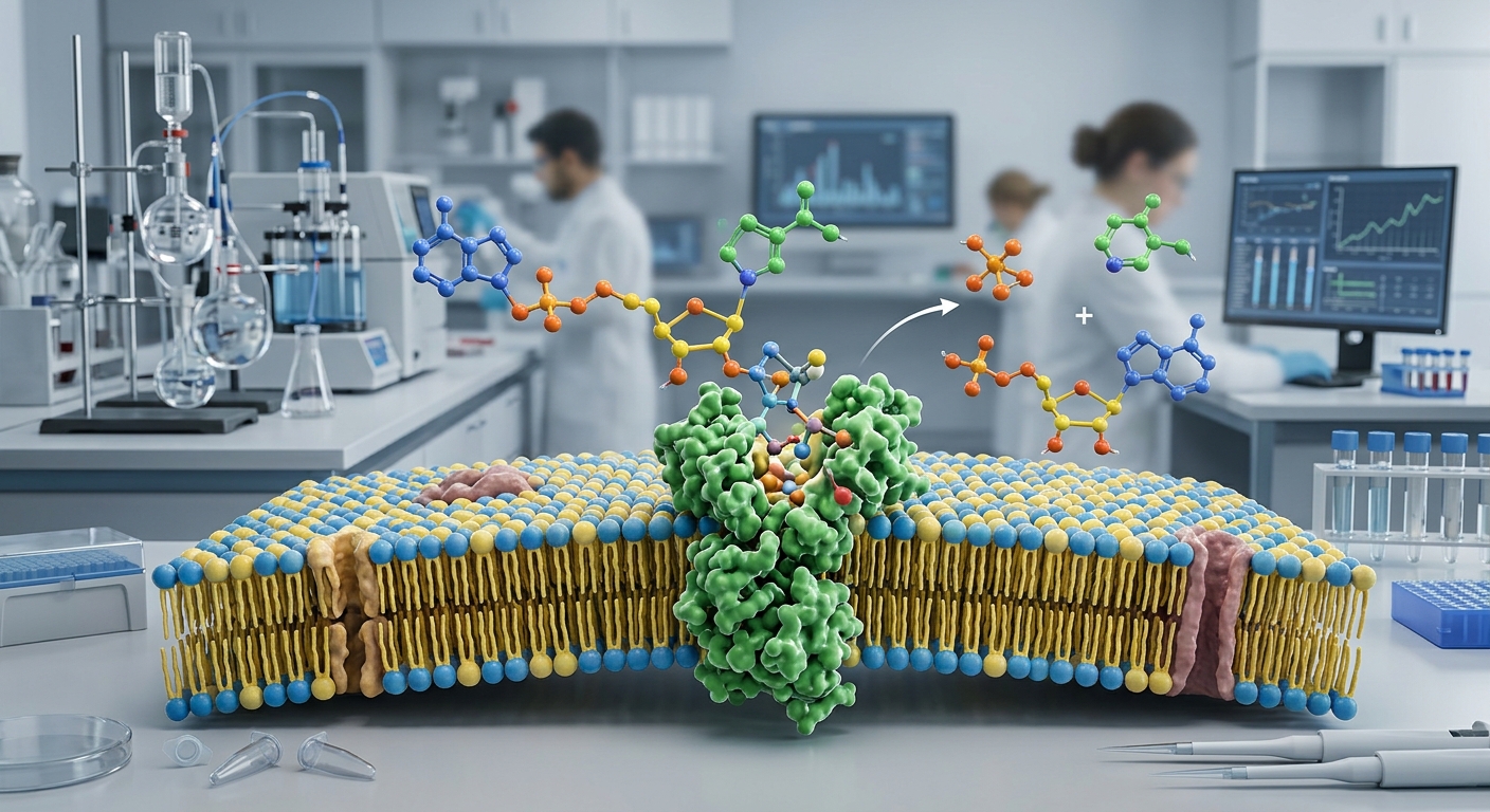
- Ecto-enzyme catabolism: Circulating and extracellular NAD+ is rapidly processed by surface enzymes, notably CD38, CD73, and ectonucleotide pyrophosphatase/phosphodiesterase 1 (ENPP1), breaking it down into smaller metabolites.
- Salvage pathway dependence: Target tissues primarily replenish intracellular NAD+ pools by importing these breakdown products (such as nicotinamide riboside and nicotinamide) and rebuilding NAD+ via intracellular salvage enzymes.
- Route-specific pharmacokinetics: Oral intact NAD+ undergoes extensive digestive and first-pass degradation, while parenteral infusions exhibit rapid clearance and metabolic transformation into intermediate metabolites rather than direct whole-cell entry.
Understanding NAD+ and Cellular Energetics
Nicotinamide adenine dinucleotide (NAD+) is a vital coenzyme present in all living cells. It exists in oxidized (NAD+) and reduced (NADH) states, acting as an essential electron carrier in fundamental biochemical pathways, including glycolysis, the tricarboxylic acid (TCA) cycle, and mitochondrial oxidative phosphorylation. Beyond its classic redox function, NAD+ serves as a consumed cosubstrate for several enzyme families, including sirtuins (SIRT1–7), poly(ADP-ribose) polymerases (PARPs), and cyclic ADP-ribose synthases (such as CD38).
Because these consuming enzymes continuously deplete the coenzyme during signaling and DNA maintenance, cells require active biosynthetic pathways to maintain intracellular homeostasis. In recent years, investigations into NAD+ bioavailability and metabolism have expanded rapidly, aiming to clarify how exogenous NAD+ is handled, degraded, and utilized by mammalian tissues.
The Intact Uptake Barrier: Molecular Structure and Transport
A central question in NAD+ pharmacology is whether intact, extracellular NAD+ can directly enter target cells. Structurally, NAD+ consists of two mononucleotides (nicotinamide mononucleotide and adenosine monophosphate) linked by a phosphoanhydride bond. This structure carries negative charges at physiological pH and exhibits substantial molecular weight (~663 Da), creating significant physicochemical barriers to passive diffusion across lipid bilayers.
While an organellar transporter known as SLC25A51 mediates intact NAD+ transport across the inner mitochondrial membrane, definitive evidence demonstrating a universal, intact plasma membrane transporter for whole-cell uptake in human tissues remains limited. Consequently, most research suggests that extracellular NAD+ cannot penetrate parenchymal cells in significant quantities without prior enzymatic breakdown.
Extracellular Metabolism: The Ecto-Enzyme Cascade
Rather than persisting as a stable intact molecule in systemic circulation or extracellular fluids, NAD+ encounters a network of membrane-bound ecto-enzymes that rapidly hydrolyze it into smaller components.
- CD38 (NAD+ Glycohydrolase): A major consumer of extracellular and circulating NAD+, CD38 cleaves the glycosidic bond of NAD+ to release nicotinamide (NAM) and ADP-ribose (ADPR) or cyclic ADPR (cADPR). CD38 expression often increases during chronic inflammation and aging, accelerating extracellular catabolism.
- ENPP1 (Ectonucleotide Pyrophosphatase/Phosphodiesterase 1): This enzyme cleaves the pyrophosphate linkage of NAD+, converting it into nicotinamide mononucleotide (NMN) and adenosine monophosphate (AMP).
- CD73 (Ecto-5′-Nucleotidase): CD73 operates downstream of ENPP1, dephosphorylating NMN to yield nicotinamide riboside (NR), while simultaneously dephosphorylating AMP into adenosine.
- Alkaline Phosphatase (ALP): Tissue-nonspecific alkaline phosphatases can also assist in the dephosphorylation of intermediate nucleotides, driving the generation of uncharged precursors.
Through this concerted enzymatic cascade, extracellular NAD+ is systematically deconstructed into smaller nucleosides and bases—primarily NR and NAM—which can then utilize dedicated equilibrative nucleoside transporters (ENTs) or organic cation transporters to enter cells.
Routes of Administration and Systemic Fate
The bioavailability of NAD+ is heavily influenced by the chosen route of administration, reflecting the susceptibility of the coenzyme to gastrointestinal, vascular, and hepatic enzymatic barriers.
Oral Ingestion and First-Pass Degradation
When administered orally, intact NAD+ encounters gastric acid and a dense brush-border enzyme system in the small intestine rich in nucleotidases and pyrophosphatases. Preclinical and clinical models demonstrate that intact dinucleotides are almost entirely degraded into nicotinamide and related metabolites before mucosal absorption. Following intestinal uptake, these metabolites travel through portal circulation and undergo extensive hepatic first-pass metabolism, where liver tissue may utilize, methylate, or release them into the broader circulation.
Intravenous Infusion Pharmacokinetics
To bypass intestinal degradation, clinical investigations have evaluated intravenous (IV) infusions of NAD+. In a landmark human pilot study conducted by Grant and colleagues (2019), researchers tracked plasma and urine metabolomes during a 6-hour constant IV infusion of 750 mg NAD+.
The study found that intact plasma NAD+ levels remained undetectable or negligible during the initial two hours of infusion, accompanied by a rapid rise in urinary excretion of methylnicotinamide (me-NAM) and adenosine metabolites. Intact NAD+ became detectable in plasma only later in the infusion window, suggesting significant initial tissue uptake or enzymatic clearance by vascular and hepatic ecto-enzymes before steady-state concentrations were reached.
Intracellular Salvage and Resynthesis
Once breakdown products cross into the intracellular compartment, target tissues utilize well-characterized biosynthetic pathways to reconstruct the NAD+ molecule:
- Nicotinamide Salvage: Nicotinamide phosphoribosyltransferase (NAMPT) catalyzes the rate-limiting conversion of NAM and 5-phosphoribosyl-1-pyrophosphate (PRPP) into NMN, which is subsequently converted to NAD+ by NMN adenylyltransferases (NMNAT1–3).
- Nicotinamide Riboside Phosphorylation: Intracellular NR is phosphorylated by nicotinamide riboside kinases (NRK1 and NRK2) to generate NMN, which then enters the standard NMNAT pathway.
Because cells rely heavily on these internal enzymes, overall NAD+ availability is dictated as much by endogenous enzyme activity (such as NAMPT and NRK levels) as by the extracellular supply of metabolites.
Safety Findings and Research Limitations
Current clinical research regarding direct NAD+ administration indicates generally favorable short-term tolerability, though rapid intravenous administration has been associated with transient flushing, chest tightness, nausea, and mild blood pressure changes. These acute effects are largely attributed to the vasoactive properties of adenosine and nicotinic acid-like intermediates formed during rapid vascular catabolism.
Several critical research limitations remain:
- Lack of human intracellular proof: While infusions raise plasma metabolite concentrations, rigorous clinical data confirming proportional increases in human parenchymal tissue NAD+ pools (such as brain or skeletal muscle) remain limited.
- Translational disparity: Animal models exhibiting drastic NAD+ recovery under experimental conditions often have artificially induced metabolic disruptions that do not reflect human physiology.
- Methylation burden: Sustained excess production of nicotinamide requires hepatic methylation (forming 1-methylnicotinamide) for excretion, raising theoretical questions regarding long-term methyl donor consumption in chronic high-dose settings.
Frequently Asked Questions
Can cells absorb intact NAD+ directly from the bloodstream?
Most mammalian cells lack plasma membrane transporters for intact NAD+. Instead, vascular and tissue-bound ecto-enzymes (such as CD38 and CD73) break circulating NAD+ down into smaller components like nicotinamide and nicotinamide riboside, which are readily transported across cell membranes and reassembled inside.
Why is oral bioavailability of intact NAD+ considered low?
The molecular size and dual negative charge of NAD+ prevent passive absorption, while intestinal enzymes and digestive acids break the molecule down into individual nucleosides and nicotinamide before it enters systemic circulation.
What is the role of CD38 in NAD+ degradation?
CD38 is a primary ecto-enzyme and cell-surface glycoprotein that hydrolyzes NAD+ into nicotinamide and ADPR. Elevated CD38 expression in inflammatory states accelerates extracellular NAD+ clearance, limiting the persistence of intact dinucleotides.
Is intravenous NAD+ approved by the FDA?
No. Intravenous NAD+ formulations are not approved by the U.S. Food and Drug Administration (FDA) for the treatment of any clinical condition, anti-aging protocol, or metabolic disorder, and remain investigational or compounded preparations.
References
- Grant, R., Berg, J., Mestayer, R., et al. (2019). A Pilot Study Investigating Changes in the Human Plasma and Urine NAD+ Metabolome During a 6 Hour Intravenous Infusion of NAD+. Frontiers in Aging Neuroscience, 11, 257.
- Gasparrini, M., Sorci, L., & Raffaelli, N. (2021). Enzymology of extracellular NAD metabolism. Cellular and Molecular Life Sciences, 78(7), 3317–3331.
- Mierzejewska, P., et al. (2021). The new insight into extracellular NAD+ degradation—the contribution of CD38 and CD73 in calcific aortic valve disease. Journal of Cellular and Molecular Medicine, 25(12), 5884–5898.
- Camacho-Pereira, J., et al. (2016). CD38 dictates age-related NAD decline and mitochondrial dysfunction through a SIRT3-dependent mechanism. Cell Metabolism, 23(6), 1127–1139.
- Luongo, T. S., et al. (2020). SLC25A51 is a mammalian mitochondrial NAD+ transporter. Nature, 588(7836), 174–179.
Research Summary
Current scientific literature demonstrates that NAD+ bioavailability and metabolism are governed by a complex ecto-enzymatic network rather than simple intact diffusion. Exogenous NAD+ is rapidly processed by enzymes such as CD38, ENPP1, and CD73 into smaller precursor molecules, which enter cells to fuel endogenous salvage pathways. While human pharmacokinetic data confirm the metabolic fate and tolerability of infused compounds, direct clinical evidence linking specific exogenous delivery forms to improved tissue outcomes remains preliminary. Unapproved for clinical use by the FDA, exogenous NAD+ remains an active subject of metabolic research.