Key Takeaways
- Engineered Structure: IGF-1 LR3 (Long Arginine 3 Insulin-Like Growth Factor-1) is an 83-amino-acid synthetic recombinant analog containing an arginine substitution at position 3 and a 13-amino-acid N-terminal peptide extension.
- IGFBP Resistance: These modifications reduce its affinity for insulin-like growth factor-binding proteins (IGFBPs) by up to 1,000-fold, allowing higher free concentrations in biological systems.
- Primary Signaling Cascades: Binding to the IGF-1 receptor (IGF-1R) stimulates receptor tyrosine kinase activity, primarily triggering the PI3K/Akt/mTOR and MAPK/ERK pathways.
- Cellular Outcomes: Downstream activation drives protein translation, cell cycle progression, myogenesis, and anti-apoptotic signaling while influencing glucose and amino acid transport.
- Research Limitations: Current findings derive almost exclusively from in vitro and animal models; IGF-1 LR3 is an unapproved investigational chemical without human clinical efficacy or safety data.
What Is IGF-1 LR3?
Insulin-like growth factor-1 Long R3 (IGF-1 LR3) is a modified recombinant peptide analog designed to overcome the rapid clearance and extensive protein-binding characteristics of endogenous human IGF-1. Naturally occurring IGF-1 consists of 70 amino acids arranged in four distinct functional domains (A, B, C, and D). In circulating human plasma, more than 98% of endogenous IGF-1 is bound to a family of six high-affinity transport proteins known as insulin-like growth factor-binding proteins (IGFBP-1 through IGFBP-6), predominantly circulating in a 150 kDa ternary complex with IGFBP-3 and an acid-labile subunit (ALS).
While IGFBPs protect endogenous peptides from proteolytic degradation, they also sequester the hormone, restricting immediate receptor engagement. IGF-1 LR3 was synthesized to manipulate this regulatory equilibrium. By combining a specific residue replacement with an N-terminal chain elongation, researchers developed an analog that largely evades binding protein sequestration while retaining high affinity for its target transmembrane receptor. Understanding the IGF-1 LR3 mechanism of action requires examining these structural alterations and the specific intracellular cascades they trigger upon receptor binding.
Structural Adaptations and IGFBP Evasion
The altered molecular behavior of IGF-1 LR3 stems from two primary modifications to the native 70-amino-acid peptide sequence:
- Glu3Arg Substitution (R3): The native glutamic acid at position 3 of the mature peptide chain is substituted with an arginine residue. Glutamic acid carries a negatively charged carboxyl side chain, whereas arginine introduces a bulky, positively charged guanidinium group. This change alters the surface charge distribution at a crucial contact point required for high-affinity association with IGFBPs.
- 13-Amino-Acid N-Terminal Extension (“Long”): A 13-residue leader sequence (Met-Phe-Pro-Ala-Met-Pro-Leu-Ser-Ser-Leu-Phe-Val-Asn) is appended to the amino terminus. This extension creates steric hindrance, physically obstructing the spatial docking geometries required by circulating binding proteins.
Together, these modifications reduce binding affinity across all six IGFBPs by 100- to 1,000-fold compared to native IGF-1. In physiological fluids and serum-supplemented cell culture media, where native IGF-1 is rapidly sequestered, IGF-1 LR3 remains predominantly in an unbound, biologically active state. Consequently, its functional in vivo half-life extends to an estimated 20 to 30 hours, compared to the roughly 15-minute half-life of free native IGF-1.
IGF-1 LR3 Mechanism of Action: Receptor Interaction
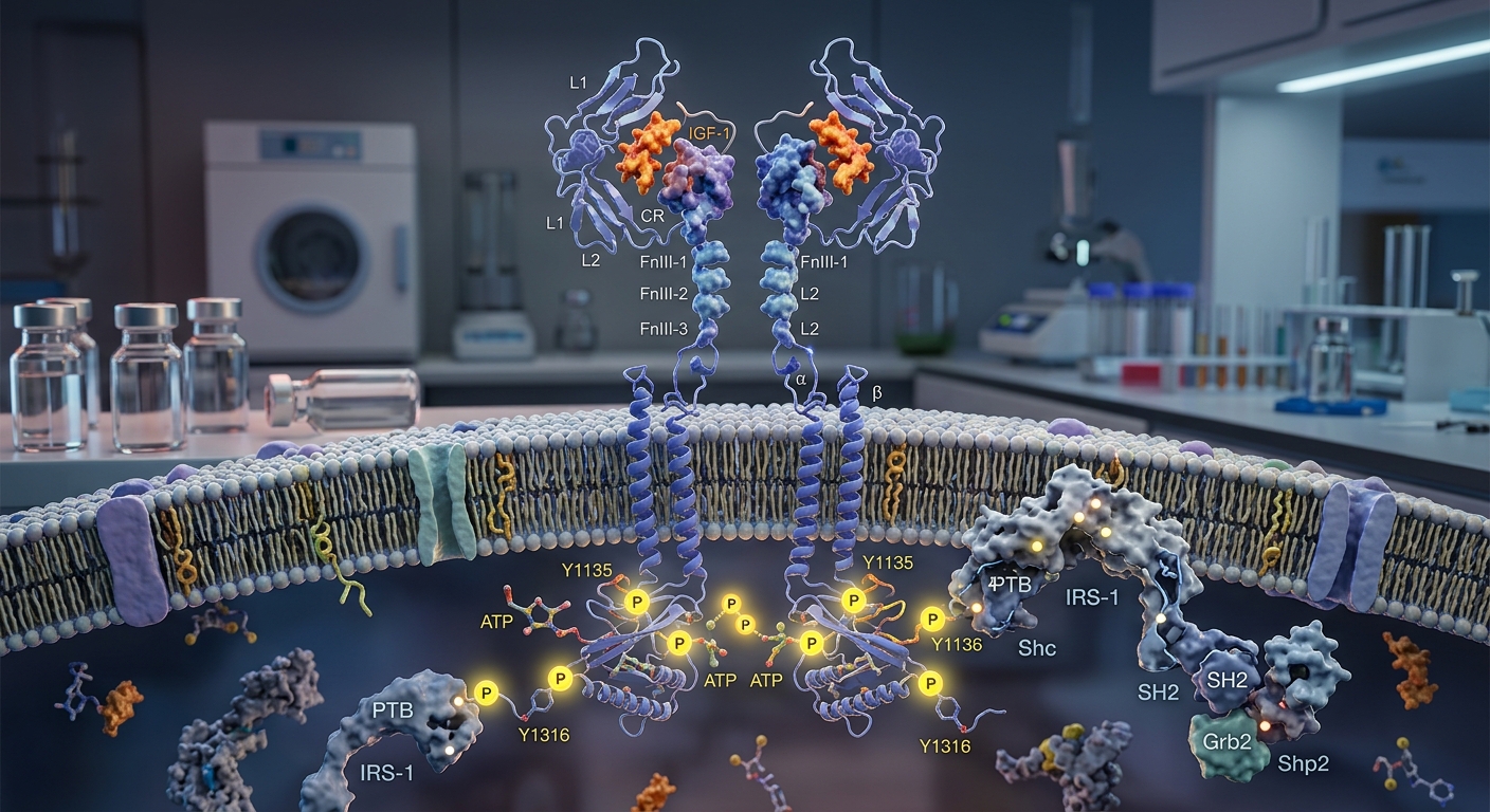
IGF-1 LR3 exerts its biological effects primarily through the type 1 insulin-like growth factor receptor (IGF-1R). The IGF-1R is a heterotetrameric transmembrane glycoprotein composed of two extracellular α-subunits that form the ligand-binding domain and two transmembrane β-subunits that possess intrinsic tyrosine kinase activity.
Receptor Binding and Transphosphorylation
When IGF-1 LR3 binds to the cysteine-rich region of the extracellular α-subunits, it induces a conformational change that brings the intracellular kinase domains of the β-subunits into proximity. This positioning stimulates trans-autophosphorylation across specific tyrosine residues within the activation loop (Tyr1131, Tyr1135, and Tyr1136). Full kinase activation creates docking phosphotyrosine motifs that recruit cytoplasmic adaptor proteins.
Adaptor Protein Recruitment
The activated receptor primarily phosphorylates two families of scaffolding molecules: Insulin Receptor Substrates (principally IRS-1 and IRS-2) and Src homology and collagen domain protein (Shc). Phosphorylated IRS proteins serve as assembly platforms that nucleate distinct multi-protein signaling complexes, branching into two principal downstream cascades: the PI3K/Akt pathway and the MAPK/ERK pathway.
Intracellular Signaling Pathways
1. The PI3K / Akt / mTOR Axis
The Phosphoinositide 3-kinase (PI3K) pathway represents the primary anabolic and cytoprotective route downstream of IGF-1R activation:
- PI3K Activation: Phosphorylated IRS-1 binds the p85 regulatory subunit of PI3K via its Src homology 2 (SH2) domains, releasing basal inhibition on the p110 catalytic subunit.
- PIP3 Generation: Active PI3K converts membrane-bound phosphatidylinositol 4,5-bisphosphate (PIP2) into phosphatidylinositol 3,4,5-trisphosphate (PIP3).
- Kinase Translocation: PIP3 recruits both Protein Kinase B (Akt) and Phosphoinositide-Dependent Kinase-1 (PDK1) to the plasma membrane via their pleckstrin homology (PH) domains. PDK1 phosphorylates Akt at Threonine-308, and mammalian target of rapamycin complex 2 (mTORC2) completes its activation by phosphorylating Serine-473.
- mTORC1 and Protein Translation: Activated Akt directly phosphorylates and inactivates the Tuberous Sclerosis Complex (TSC1/TSC2), relieving inhibition on Rheb and promoting mammalian target of rapamycin complex 1 (mTORC1) assembly. mTORC1 phosphorylates downstream effectors p70S6 kinase (p70S6K) and eukaryotic initiation factor 4E-binding protein 1 (4E-BP1), initiating ribosomal biogenesis and peptide translation.
- Cell Survival Signaling: Akt phosphorylates and inactivates the pro-apoptotic protein Bad and members of the Forkhead box O (FoxO) transcription factor family. FoxO phosphorylation sequesters these proteins in the cytoplasm, preventing the transcription of apoptotic and proteolytic genes, including the muscle-specific E3 ubiquitin ligases atrogin-1 and MuRF1.
2. The MAPK / ERK Signaling Axis
Parallel to PI3K activation, the phosphorylated β-subunits of IGF-1R and phosphorylated Shc recruit the adaptor protein Grb2 complexed with Son of Sevenless (SOS), a guanine nucleotide exchange factor:
- SOS promotes the exchange of GDP for GTP on the membrane-anchored G-protein Ras.
- Active Ras-GTP recruits and activates the serine/threonine kinase Raf.
- Raf initiates a sequential phosphorylation cascade, activating Mitogen-Activated Protein Kinase Kinase (MEK1/2), which directly phosphorylates Extracellular Signal-Regulated Kinases 1 and 2 (ERK1/2).
- Phosphorylated ERK1/2 translocates to the nucleus to activate key transcription factors, such as c-Jun, c-Fos, and Elk-1, regulating cell cycle progression, proliferation, and differentiation.
Cellular Effects in Preclinical Models
Because IGF-1 LR3 maintains continuous receptor engagement without rapid IGFBP neutralization, it produces pronounced biological responses in laboratory settings:
- Myogenesis and Satellite Cell Dynamics: In cell models (such as murine C2C12 and L6 myoblasts), sustained IGF-1R signaling stimulates both myoblast proliferation via MAPK/ERK and terminal differentiation into myotubes via PI3K/Akt.
- Nutrient Transport: Akt activation promotes the translocation of glucose transporter type 4 (GLUT4) vesicles to the cell membrane, facilitating insulin-independent glucose uptake into skeletal myocytes and adipocytes.
- Cellular Longevity: Prolonged suppression of caspase activation and inhibition of pro-apoptotic Bcl-2 family members promote survival under serum-deprived or metabolically stressed culture conditions.
Safety, Cross-Reactivity, and Research Limitations
Despite significant preclinical utility, several important biological limitations and physiological risks exist regarding this analog:
- Insulin Receptor Cross-Reactivity: At elevated concentrations, IGF-1 LR3 cross-reacts with homodimeric insulin receptors (IR) and hybrid IGF-1R/IR complexes. In animal studies, continuous receptor occupancy can cause profound hypoglycemia through excessive peripheral glucose clearance and suppression of hepatic gluconeogenesis.
- Mitogenic and Neoplastic Concerns: Sustained activation of the IGF-1R/Akt/ERK axes inhibits apoptosis and accelerates cellular proliferation, raising concerns in preclinical oncology regarding the promotion of abnormal cellular growth and neoplasia.
- Pituitary Feedback Disruption: Systemic administration in animal models suppresses endogenous growth hormone (GH) secretion and hepatic IGF-1 synthesis through negative feedback on the hypothalamic-pituitary-somatotropic axis.
- Absence of Human Trials: The pharmacokinetic advantages observed in laboratory cell lines have not been evaluated in controlled human clinical trials. There are no robust safety, dosing, or long-term toxicology data for human populations.
Regulatory Status
IGF-1 LR3 is an unapproved investigational compound. It is not approved by the U.S. Food and Drug Administration (FDA), the European Medicines Agency (EMA), or any other international health agency for diagnostic, therapeutic, or dietary use. While recombinant human IGF-1 (mecasermin) holds approval for specific pediatric growth disorders, modified variants like IGF-1 LR3 remain restricted solely to laboratory and in vitro scientific research.
Frequently Asked Questions
How does IGF-1 LR3 differ from endogenous IGF-1?
Endogenous IGF-1 contains 70 amino acids and binds tightly to circulating IGF-binding proteins (IGFBPs). IGF-1 LR3 has 83 amino acids, featuring an arginine substitution at position 3 and a 13-amino-acid N-terminal leader sequence. These modifications reduce its affinity for IGFBPs, keeping the analog in a free, receptor-active state for a substantially longer duration.
Which receptors does IGF-1 LR3 bind?
IGF-1 LR3 functions as a high-affinity agonist at the type 1 IGF receptor (IGF-1R). It also exhibits lower-affinity cross-reactivity with insulin receptors (IR) and hybrid IGF-1R/IR heterodimers. Unlike native IGF-1, it shows reduced binding to the mannose-6-phosphate/IGF-2 clearance receptor.
Why is IGF-1 LR3 commonly used in cell culture?
In serum-containing cell cultures, endogenous IGFBPs sequester standard recombinant IGF-1, attenuating its signaling efficiency. IGF-1 LR3 resists this sequestration, allowing researchers to maintain steady IGF-1R activation at lower nanomolar concentrations without frequent replenishment.
What are the primary signaling pathways activated by IGF-1 LR3?
IGF-1 LR3 primarily activates two pathways: the PI3K/Akt/mTOR pathway (which governs protein synthesis, metabolic actions, and cell survival) and the Ras/Raf/MEK/ERK pathway (which regulates gene expression and cell proliferation).
References
- Tomas FM, Lemmey AB, Read LC, Ballard FJ. Superior potency of infused IGF-I analogues which bind poorly to IGF-binding proteins is maintained when administered by injection. J Endocrinol. 1996;150(1):77-84.
- Mohan S, Baylink DJ. IGF-binding proteins are multifunctional and act via IGF-dependent and -independent mechanisms. J Endocrinol. 2002;175(1):19-31.
- Assefa B, et al. Insulin-Like Growth Factor (IGF) Binding Protein-2, Independently of IGF-1, Induces GLUT-4 Translocation and Glucose Uptake in 3T3-L1 Adipocytes. Oxid Med Cell Longev. 2017;2017:3035184.
- Parrado Fernandez C, et al. Intranasal Long R3 insulin-like growth factor-1 treatment promotes amyloid plaque remodeling in cerebral cortex, but fails to preserve cognitive function in male 5XFAD mice. Brain Commun. 2024;6(1):fcae017.
Research Summary
Current published literature demonstrates that IGF-1 LR3 functions as a long-acting agonist of the IGF-1 receptor, relying on structural alterations (Glu3Arg and a 13-residue N-terminal extension) to evade IGFBP binding and prolong downstream PI3K/Akt and MAPK/ERK signaling. However, empirical evidence remains limited to in vitro and non-human animal models. There are no completed, peer-reviewed human clinical trials validating its therapeutic efficacy, pharmacokinetics, or safety profile in humans. IGF-1 LR3 remains an unapproved research compound restricted strictly to laboratory investigation.