Key Takeaways
- Sequence and Origin: KPV is a synthetic tripeptide consisting of Lysine-Proline-Valine (Lys-Pro-Val), representing the carboxy-terminal residues 11–13 of alpha-melanocyte-stimulating hormone (alpha-MSH).
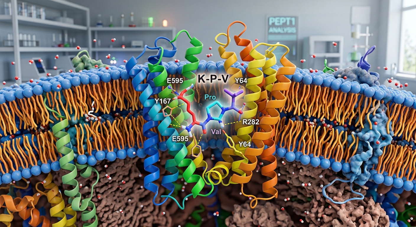
- Transport Mechanism: KPV enters target cells through the proton-coupled oligopeptide transporter PepT1 (encoded by SLC15A1), an apical transporter expressed in the small intestine and aberrantly upregulated in inflamed colonic tissue.
- Signaling Inhibition: Inside epithelial and immune cells, KPV impedes the activation of nuclear factor kappa B (NF-κB) and mitogen-activated protein kinase (MAPK) cascades, curbing downstream proinflammatory cytokine production.
- Preclinical Colitis Models: In murine models of chemical colitis (DSS and TNBS), orally administered KPV has demonstrated marked reductions in mucosal ulceration, myeloperoxidase activity, and histological injury scores.
- Translational Status: Despite robust laboratory findings, KPV lacks human clinical trials, has not received regulatory approval from the U.S. Food and Drug Administration (FDA), and remains restricted to experimental research.
What Is KPV? Molecular Profile and Origins
KPV is an ultra-short tripeptide with the primary amino acid sequence L-lysyl-L-prolyl-L-valine. Structurally, it represents the C-terminal tripeptide fragment of alpha-melanocyte-stimulating hormone (alpha-MSH), an endogenous peptide hormone cleaved from the proopiomelanocortin (POMC) precursor. While full-length alpha-MSH exerts systemic endocrine effects, including skin pigmentation via the melanocortin-1 receptor (MC1R), investigative attention turned to smaller fragments capable of retaining anti-inflammatory properties without triggering receptor-driven pigmentary changes.
Preclinical investigations revealed that the tripeptide fragment preserves significant anti-inflammatory signaling capability. Unlike its full-length parent hormone, KPV operates predominantly independent of classical melanocortin receptor activation. With a low molecular weight of 342.43 g/mol, the peptide presents minimal steric resistance, allowing it to behave as an efficient substrate for specialized nutrient and peptide transport systems across epithelial surfaces.
The PepT1 Transport Mechanism in Intestinal Epithelium
A central question in peptide pharmacology is how small, hydrophilic molecules traverse mucosal membranes to exert intracellular effects. For KPV, uptake in the gastrointestinal tract is mediated primarily by the human peptide transporter 1 (hPepT1, gene symbol SLC15A1). PepT1 is a high-capacity, low-affinity, proton-dependent symporter whose canonical physiological role is the absorption of dietary dipeptides and tripeptides across the brush-border membrane of the small intestine.
Under healthy physiological conditions, PepT1 is abundantly expressed in the duodenum, jejunum, and ileum, but is virtually absent or expressed at negligible levels in the healthy human colon. However, during active intestinal inflammation—such as in ulcerative colitis or Crohn’s disease—PepT1 undergoes marked ectopic upregulation in the colonic epithelium and infiltrating immune cells, including macrophages and T lymphocytes. This disease-induced upregulation creates a localized cellular doorway specifically in areas of mucosal injury.
A landmark investigation published in Gastroenterology by Dalmasso and colleagues (2008) demonstrated that KPV acts as a direct substrate for PepT1. Competition assays using radiolabeled PepT1 substrates confirmed that KPV enters intestinal epithelial cell lines (such as Caco2-BBE and HT-29) and Jurkat T cells in a saturable, carrier-mediated fashion. Because colonic inflammation drives PepT1 expression, the transporter functions as an opportunistic gateway, enabling KPV to concentrate directly within the inflamed tissue microenvironment.
Intracellular Actions: NF-κB and MAPK Pathway Inhibition
Once transported across the plasma membrane, KPV interferes with core transcriptional programs that govern intestinal inflammation. The best-characterized target of intracellular KPV is the nuclear factor kappa B (NF-κB) signaling network. Under basal conditions, NF-κB dimers (such as p65/p50) remain sequestered in the cytoplasm bound to the inhibitory protein IκBα. In response to proinflammatory stimuli—such as tumor necrosis factor-alpha (TNF-α) or interleukin-1 beta (IL-1β)—the IκB kinase (IKK) complex phosphorylates IκBα, triggering its proteasomal degradation and allowing free NF-κB dimers to translocate to the nucleus.
In human intestinal epithelial cell models, nanomolar concentrations of KPV significantly suppress NF-κB-driven luciferase reporter activity and prevent the degradation of IκBα. By halting nuclear accumulation of p65, KPV inhibits the transcriptional activation of critical inflammatory mediators, including interleukin-8 (IL-8), TNF-α, and mucosal chemokines. Complementary research in related epithelial models suggests that C-terminal alpha-MSH peptides can directly interact with nuclear transport proteins (such as importin-alpha3), competitively impeding nuclear access of the p65 subunit.
In addition to NF-κB suppression, KPV dampens activation within the mitogen-activated protein kinase (MAPK) superfamily. Preclinical assays demonstrate diminished phosphorylation of extracellular signal-regulated kinases (ERK1/2) and p38 MAPK following KPV administration. By intercepting both NF-κB and MAPK cascades simultaneously, KPV disrupts the primary signaling hubs responsible for chronic mucosal damage.
Preclinical Evidence in Rodent Colitis Models
The biological efficacy of KPV in intestinal inflammation has been documented across distinct experimental animal models of inflammatory bowel disease (IBD):
- Dextran Sulfate Sodium (DSS) Colitis: The DSS model induces toxic injury to the colonic epithelial monolayer, driving mucosal erosions, hematochezia, and granulocyte infiltration that mimic acute flares of ulcerative colitis. In mice receiving oral KPV via drinking water, researchers observed a significant attenuation of weight loss, reduced disease activity indices, and markedly diminished colonic myeloperoxidase (MPO) activity—a primary histological marker of neutrophil infiltration.
- Trinitrobenzene Sulfonic Acid (TNBS) Colitis: TNBS induces a transmural, Th1/Th17-mediated inflammatory response that approximates Crohn’s disease pathology. In TNBS-challenged mice, KPV administration maintained colonic wall thickness, prevented severe mucosal ulceration, and suppressed mRNA expression of proinflammatory cytokines (including TNF-α and IL-1β) within colonic tissue extracts.
- Colitis-Associated Carcinogenesis (CAC): Chronic intestinal inflammation dramatically increases the risk of neoplastic transformation. A follow-up study by Viennois et al. (2016) in Cellular and Molecular Gastroenterology and Hepatology investigated KPV in an azoxymethane (AOM)/DSS murine model. KPV administration significantly decreased colonic tumor incidence and total tumor burden. Crucially, this protective benefit was completely absent in PepT1-knockout mice, confirming that the therapeutic actions of KPV depend strictly upon functional PepT1 transport.
Formulation and Drug Delivery Challenges
Despite promising preclinical efficacy, the translational viability of native KPV faces notable pharmacokinetic hurdles. As an unprotected linear tripeptide, KPV is susceptible to rapid enzymatic degradation by brush-border peptidases, gastric acid, and systemic aminopeptidases. Oral administration of native peptides often results in poor bioavailability due to premature cleavage before reaching the distal colon.
To overcome this limitation, bioengineers have designed targeted nanocarrier systems. In murine colitis studies, researchers synthesized hyaluronic acid-functionalized nanoparticles encapsulated with KPV (HA-KPV-NPs). Hyaluronic acid binds with high affinity to CD44 receptors, which are heavily upregulated on inflamed colonic epithelial cells. These nanoparticle systems protected KPV from premature gastrointestinal degradation and enabled mucosal healing at dosages several thousand-fold lower than unencapsulated free peptide, illustrating that successful clinical translation will likely rely on advanced colon-targeted drug delivery platforms.
Evidence Gaps and Translational Limitations
While the laboratory literature is substantial, significant evidentiary gaps restrict the interpretation of these findings:
- Absence of Human Trials: To date, there are no published randomized controlled trials or Phase I/II safety studies assessing KPV in human patients with ulcerative colitis or Crohn’s disease. Findings in rodent models cannot guarantee efficacy, safety, or appropriate dosing in clinical populations.
- Pharmacokinetic Uncertainties: The metabolic half-life, systemic clearance, tissue distribution, and degradation products of KPV in humans remain largely undefined.
- Heterogeneity of Human IBD: Chemically induced rodent colitis models (DSS, TNBS) model acute mucosal insult rather than the complex, relapsing, polygenic pathophysiology characteristic of human inflammatory bowel disease.
Regulatory Status
KPV is not approved by the U.S. Food and Drug Administration (FDA) or the European Medicines Agency (EMA) for the prevention, diagnosis, or treatment of any medical condition. It has not been evaluated in formal new drug application (NDA) pathways. In regulatory classifications, KPV is designated strictly as an investigational research chemical intended for in vitro and laboratory animal research. It is not an approved dietary supplement, prescription drug, or authorized compound for clinical use.
Frequently Asked Questions
Does KPV bind to melanocortin receptors?
Unlike its parent hormone alpha-MSH, KPV does not exhibit meaningful agonist activity at melanocortin receptors MC1R, MC3R, MC4R, or MC5R. Its anti-inflammatory actions in intestinal models occur through intracellular mechanisms following PepT1-mediated transport, avoiding the pigmentary or endocrine effects associated with melanocortin receptor activation.
Why is PepT1 critical to KPV research?
PepT1 (SLC15A1) serves as the primary transporter responsible for moving KPV across cell membranes into the cytoplasm of intestinal epithelial and immune cells. Genetic knockout studies confirm that without functional PepT1, KPV fails to enter target cells and cannot suppress mucosal inflammation or colitis-associated tumorigenesis.
Can KPV replace standard therapies for ulcerative colitis?
No. Standard therapies for ulcerative colitis—such as 5-aminosalicylates, corticosteroids, immunomodulators, and biologic agents—are backed by robust clinical trial data and established safety profiles. KPV remains an unapproved experimental compound supported solely by preclinical cell and animal experiments.
Research Summary
Preclinical studies demonstrate that the alpha-MSH-derived tripeptide KPV possesses distinct anti-inflammatory properties relevant to intestinal pathology. Transported into cells via the proton-coupled oligopeptide transporter PepT1 (which is selectively upregulated during active colonic inflammation), KPV inhibits the NF-κB and MAPK signaling pathways to suppress cytokine transcription. In rodent models of acute chemical colitis and colitis-associated carcinogenesis, oral KPV has consistently reduced mucosal damage and tumor progression in a PepT1-dependent manner. However, the entirety of published evidence remains strictly preclinical. KPV has not been tested in clinical trials, lacks FDA approval, and requires substantial pharmacokinetic validation before human therapeutic relevance can be determined.
References
- Dalmasso G, Charrier-Hisamuddin L, Nguyen HT, Yan Y, Sitaraman S, Merlin D. PepT1-mediated tripeptide KPV uptake reduces intestinal inflammation. Gastroenterology. 2008;134(1):166-178. doi:10.1053/j.gastro.2007.10.026
- Viennois E, Xiao B, Ayyadurai S, Wang L, Han MK, Zhang M, Merlin D. Critical role of PepT1 in promoting colitis-associated cancer and therapeutic benefits of the anti-inflammatory PepT1-mediated tripeptide KPV in a murine model. Cell Mol Gastroenterol Hepatol. 2016;2(3):340-357. doi:10.1016/j.jcmgh.2016.01.006
- Dalmasso G, et al. PepT1-Mediated Tripeptide KPV Uptake Reduces Intestinal Inflammation (Full Text). PubMed Central. PMCID: PMC2431115.