Key Takeaways
- Primary Central Antioxidant: Glutathione (GSH) is the central nervous system’s principal endogenous antioxidant, responsible for maintaining redox balance, neutralizing reactive oxygen species, and preserving mitochondrial integrity.
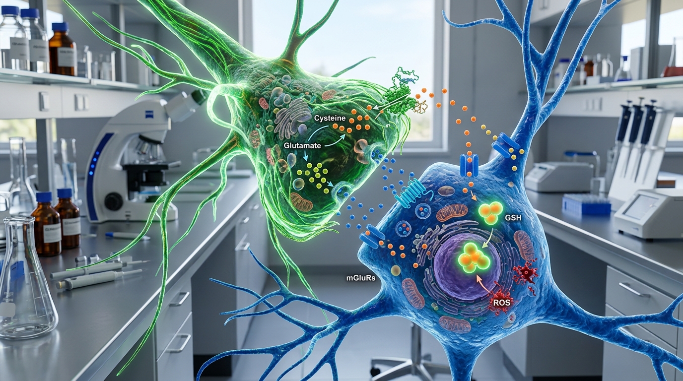
- Astrocyte-Neuron Cooperation: Neurons have a limited capacity to synthesize glutathione autonomously and depend on neighboring astrocytes to supply precursor amino acids, notably cysteine.
- Delivery Challenges: Intact exogenous glutathione exhibits poor oral bioavailability and limited transport across the blood-brain barrier, making central nervous system delivery a major research hurdle.
- Human Clinical Evidence: While preclinical rodent and cellular models show neuroprotective effects, human clinical trials examining intravenous and intranasal administration have yielded mixed and preliminary results.
The Role of Glutathione in Neural Redox Balance
The human brain accounts for approximately 20% of resting metabolic energy consumption despite representing only 2% of total body mass. This intense metabolic activity generates substantial baseline levels of reactive oxygen species (ROS) and reactive nitrogen species (RNS) during mitochondrial oxidative phosphorylation. Neural tissue is uniquely susceptible to oxidative stress due to its high lipid content—particularly polyunsaturated fatty acids prone to lipid peroxidation—and elevated concentrations of redox-active transition metals such as iron.
Glutathione is a tripeptide composed of glutamate, cysteine, and glycine (gamma-L-glutamyl-L-cysteinylglycine). In its reduced monomeric form (GSH), it acts as a primary cellular nucleophile and free-radical scavenger. Glutathione peroxidase (GPx) utilizes GSH as an electron donor to reduce hydrogen peroxide and lipid hydroperoxides into water and lipid alcohols, producing glutathione disulfide (GSSG) in the process. The enzyme glutathione reductase (GR) subsequently regenerates GSH from GSSG using NADPH as a reducing cofactor, maintaining a high intracellular GSH:GSSG ratio under physiological conditions.
Astrocyte-Neuron Metabolic Coupling and Synthesis
Glutathione synthesis in neural tissue involves a two-step ATP-dependent enzymatic cascade. First, glutamate-cysteine ligase (GCL)—composed of catalytic (GCLC) and modifier (GCLM) subunits—catalyzes the rate-limiting formation of gamma-glutamylcysteine from L-glutamate and L-cysteine. Second, glutathione synthetase adds glycine to complete the tripeptide.
Neurons and astrocytes exhibit distinct metabolic roles in sustaining brain glutathione concentrations:
- Astrocytic Synthesis: Astrocytes synthesize high concentrations of GSH and release it into the extracellular space via the multidrug resistance protein 1 (MRP1) transporter.
- Extracellular Cleavage: The astroglial ectoenzyme gamma-glutamyl transpeptidase (GGT) and aminopeptidase N cleave extracellular GSH into dipeptides and constituent amino acids, primarily Cys-Gly and free cysteine.
- Neuronal Uptake: Neurons import these precursor fragments via excitatory amino acid transporters (EAATs) and dipeptide transporters, using them to synthesize intra-neuronal GSH.
Disruptions in this astrocyte-neuron metabolic axis can compromise neuronal antioxidant defenses and increase vulnerability to excitotoxicity and mitochondrial dysfunction.
Preclinical Evidence in Neurodegenerative Models
In vitro and animal models have consistently demonstrated that depletion of brain glutathione correlates with accelerated neuronal damage, mitochondrial complex I inhibition, and increased sensitivity to oxidative stressors.
Parkinson’s Disease Models
Post-mortem analyses of human brain tissue have documented selective reductions in total glutathione within the substantia nigra pars compacta in early stages of Parkinson’s disease. In rodent models using neurotoxins such as 6-hydroxydopamine (6-OHDA) or MPTP, experimental depletion of GSH exacerbates dopaminergic neuron degeneration. Conversely, genetic overexpression of GCLC or administration of glutathione precursors (such as N-acetylcysteine) has shown protective effects against oxidative damage in these animal models.
Ischemia and Reperfusion Injury
Cerebral ischemia-reperfusion models demonstrate that a transient loss of blood flow followed by reoxygenation produces a burst of ROS that rapidly depletes endogenous GSH. Preclinical investigations into GSH-replenishing agents, including glutathione ethyl ester and synthetic glutathione mimetics, show reductions in infarct volume and lipid peroxidation biomarkers in rodent transient middle cerebral artery occlusion (tMCAO) paradigms.
Clinical Research and Pharmacokinetic Barriers
Translating preclinical findings into effective human neuroprotective strategies has encountered major obstacles, predominantly involving delivery pharmacokinetics and rapid clearance.
The Blood-Brain Barrier (BBB)
Systemically administered intact glutathione has poor permeability across the blood-brain barrier. The BBB endothelial cells express gamma-glutamyl transferase, which degrades intact circulatory peptide into individual amino acids before significant direct transport into the brain parenchyma can occur. Consequently, oral and intravenous administration produce minimal increases in brain GSH levels in clinical imaging studies.
Intravenous Clinical Trials
Several early open-label and small randomized controlled trials evaluated intravenous glutathione infusions in individuals with Parkinson’s disease. While initial open-label reports suggested transient symptomatic improvements, subsequent randomized, double-blind, placebo-controlled trials failed to establish a statistically significant therapeutic difference between intravenous glutathione and placebo on validated motor rating scales over multi-week protocols.
Intranasal Administration
To bypass the BBB, researchers have investigated intranasal glutathione delivery via the olfactory and trigeminal neural pathways directly into the central nervous system. Phase I and Phase II randomized trials assessing intranasal glutathione in neurodegenerative cohorts have shown that the route is generally tolerated and transiently increases baseline GSH concentrations measured by magnetic resonance spectroscopy (MRS). However, these studies have been limited by small sample sizes, short study durations, and lack of consistent clinical efficacy endpoints.
Current Research Directions and Delivery Strategies
To overcome pharmacokinetic limitations, research is exploring alternative delivery platforms and metabolic strategies:
- Glutathione Precursors: Investigating agents like N-acetylcysteine (NAC) and NAC amide, which cross biological membranes more readily to provide the rate-limiting cysteine precursor for de novo synthesis.
- Nanoparticle Encapsulation: Developing liposomal, polymeric, and solid lipid nanoparticles designed to protect the tripeptide from systemic degradation and facilitate transcytosis across the BBB.
- Nrf2 Pathway Activators: Studying small molecules that upregulate Nuclear factor erythroid 2-related factor 2 (Nrf2), the master transcriptional regulator of endogenous antioxidant response elements (ARE), which increases expression of GCL and GPx enzymes.
Frequently Asked Questions
Can oral glutathione supplements increase brain glutathione levels?
Oral intact glutathione undergoes significant degradation by digestive peptidases and hepatic first-pass metabolism, resulting in very low intact systemic bioavailability. Current scientific evidence indicates that standard oral glutathione does not significantly raise central nervous system glutathione concentrations.
How is brain glutathione measured in clinical studies?
In living human subjects, brain glutathione levels are non-invasively measured using proton magnetic resonance spectroscopy (1H-MRS). Special spectral editing techniques, such as MEGA-PRESS, are utilized to distinguish the glutathione signal from more abundant brain metabolites such as GABA and glutamate.
Is glutathione approved by regulatory agencies for neuroprotection?
No. Glutathione is not approved by the U.S. Food and Drug Administration (FDA) or other major regulatory agencies for the treatment or prevention of any neurodegenerative disease or neurological disorder. Its use in this domain remains strictly investigational.
Research Summary
Preclinical research firmly establishes glutathione as a critical mediator of neural redox balance and neuronal survival under oxidative stress. However, translation to clinical efficacy in neurodegenerative and cerebrovascular conditions remains unproven. The primary limitations include poor blood-brain barrier transport, rapid degradation of systemic intact peptide, and inconclusive clinical trial outcomes. This review synthesizes general domain knowledge; specific experimental protocols and clinical trials should be independently cross-referenced via peer-reviewed literature and PubMed.