Key Takeaways
- Distinct Mode of Death: Ferroptosis is an iron-dependent, non-apoptotic form of regulated cell death characterized by catastrophic lipid peroxidation in cellular membranes.
- Core Defense Mechanism: Reduced glutathione (GSH) is the indispensable electron donor for glutathione peroxidase 4 (GPX4), the primary enzyme that converts toxic lipid hydroperoxides into non-toxic lipid alcohols.
- The System Xc− Connection: Cellular GSH synthesis relies heavily on the cystine/glutamate antiporter (System Xc−, composed of SLC7A11 and SLC3A2); inhibiting this transporter depletes intracellular cysteine and GSH, triggering ferroptosis.
- Therapeutic Paradox: In preclinical oncology, researchers actively induce ferroptosis by depleting GSH to eliminate drug-resistant tumor cells. Conversely, in neurodegenerative and ischemic injury models, maintaining GSH levels protects vulnerable tissues.
What Is Ferroptosis and Why Does Glutathione Matter?
Ferroptosis is a form of regulated cell death (RCD) first formally characterized in 2012 by Dr. Brent R. Stockwell’s laboratory. Unlike apoptosis, which depends on caspase activation and cellular packaging into apoptotic bodies, or necroptosis, which relies on receptor-interacting protein kinases, ferroptosis is driven entirely by biochemical failure: the lethal accumulation of iron-dependent lipid reactive oxygen species (ROS) within membrane phospholipids.
At the center of cellular defense against ferroptosis is glutathione (GSH), a tripeptide composed of glutamate, cysteine, and glycine. GSH is the most abundant non-protein thiol in eukaryotic cells, maintaining redox homeostasis and neutralizing oxidative insults. When GSH levels plummet or its downstream enzymatic partners are inactivated, cellular membranes undergo unchecked radical chain reactions, rupturing the phospholipid bilayer and executing ferroptotic lysis.
The System Xc− / GSH / GPX4 Axis
The primary barrier against ferroptotic cell death is the System Xc− / GSH / GPX4 axis. This metabolic pathway links nutrient uptake directly to membrane lipid preservation through three tightly integrated steps:
- Cystine Import via System Xc−: System Xc− is a heterodimeric amino acid transporter spanning the plasma membrane, consisting of the functional subunit SLC7A11 and the regulatory chaperone SLC3A2. It imports extracellular cystine in exchange for intracellular glutamate. Once imported, cystine is rapidly reduced to cysteine, the rate-limiting amino acid for de novo GSH synthesis.
- Glutathione Biosynthesis: Intracellular cysteine is combined with glutamate by glutamate-cysteine ligase (GCL), and then joined with glycine by glutathione synthetase (GSS), yielding reduced glutathione (GSH).
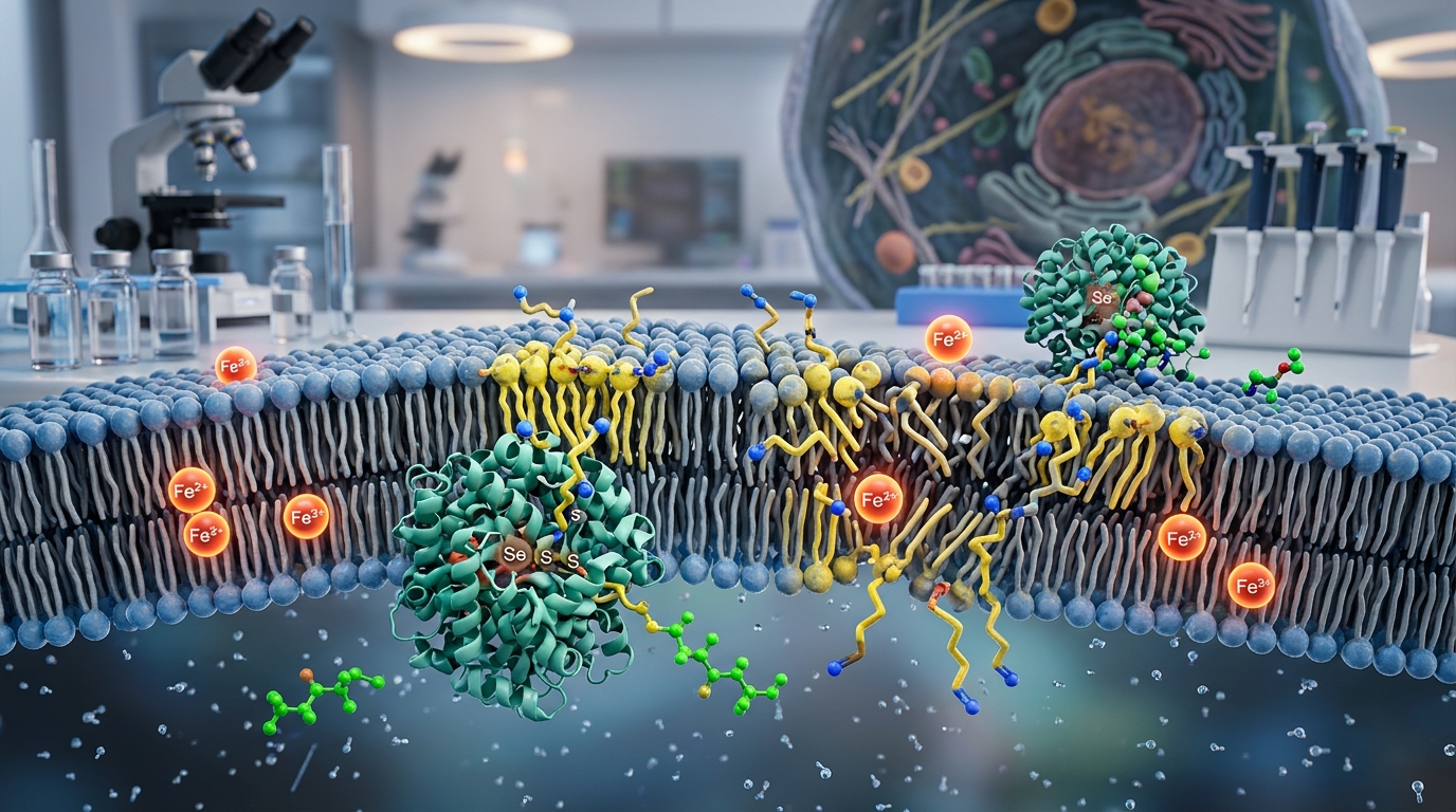
- GPX4 Catalytic Reduction: Glutathione peroxidase 4 (GPX4) is a unique selenoprotein that directly reduces complex phospholipid hydroperoxides (PLOOH) to their corresponding non-toxic phospholipid alcohols (PLOH). In this catalytic cycle, GPX4 utilizes two molecules of GSH as electron donors, converting them into oxidized glutathione disulfide (GSSG).
Mechanisms of Lipid Peroxidation and Iron Toxicity
Polyunsaturated fatty acids (PUFAs), particularly arachidonic acid and adrenic acid esterified into phosphatidylethanolamines (PE), are exceptionally vulnerable to oxidation. Enzymes such as acyl-CoA synthetase long-chain family member 4 (ACSL4) and lysophosphatidylcholine acyltransferase 3 (LPCAT3) incorporate these oxidizable PUFAs into membrane phospholipids.
When cellular iron metabolism yields an excess of the labile iron pool (Fe2+), iron catalyzes the Fenton reaction with endogenous peroxides. This generates highly reactive hydroxyl or alkoxyl radicals that abstract hydrogen atoms from PUFA carbon chains, instigating a self-propagating peroxidation cascade. Under normal physiological conditions, GPX4 consumes GSH to arrest this cascade immediately. If GSH is depleted, GPX4 loses its reducing substrate, allowing membrane peroxides to accumulate until structural integrity fails.
Pharmacological Modulation in Laboratory Research
Preclinical research frequently uses selective small molecules to dissect the mechanics of glutathione and ferroptosis. These compounds are categorized based on their specific molecular targets:
- Class I Inducers (GSH Depletion): Compounds such as erastin and sulfasalazine directly inhibit System Xc−, preventing cystine uptake and starving the cell of cysteine. Similarly, buthionine sulfoximine (BSO) inhibits glutamate-cysteine ligase, directly halting GSH synthesis. Both approaches trigger ferroptosis upstream of GPX4.
- Class II Inducers (GPX4 Inactivation): Molecules such as RSL3 and ML162 bind covalently to the selenocysteine active site of GPX4, inactivating the enzyme directly without requiring immediate GSH depletion.
- Ferroptosis Inhibitors: Lipophilic radical-trapping antioxidants such as ferrostatin-1 (Fer-1) and liproxstatin-1, as well as iron chelators like deferoxamine (DFO), suppress lipid peroxidation and rescue cells even when GSH is depleted.
Preclinical Applications: A Dual Biological Role
Because glutathione-dependent ferroptosis regulation governs cell survival under oxidative stress, it represents a dual-edged sword in biomedical research:
Oncology: Exploiting Metabolic Vulnerabilities
Many therapy-resistant cancer cell lines—particularly those undergoing epithelial-mesenchymal transition (EMT) or carrying oncogenic mutations—exhibit heightened reliance on SLC7A11 and GPX4 for survival. In laboratory models, depleting GSH or blocking GPX4 selectively triggers ferroptotic destruction of chemotherapy-resistant tumors, providing an experimental strategy to bypass classical apoptosis resistance.
Ischemic Injury and Neurodegeneration: Halting Pathological Loss
Conversely, excessive ferroptosis contributes significantly to pathological tissue loss. In preclinical models of stroke, myocardial ischemia-reperfusion injury, Parkinson’s disease, and Alzheimer’s disease, localized GSH depletion and iron accumulation drive neuronal and cardiomyocyte death. In these contexts, researchers explore strategies to maintain intracellular GSH pools or administer lipid radical scavengers to preserve tissue function.
Limitations and Evidence Gaps
While the relationship between glutathione and ferroptosis is supported by extensive in vitro and animal models, critical gaps remain:
- Parallel Antioxidant Pathways: GSH and GPX4 do not act in complete isolation. Alternative ferroptosis suppressor pathways—such as the FSP1-CoQ10 axis, the DHODH pathway in mitochondria, and the GCH1-BH4 system—can compensate for GSH deficiency in certain cell types.
- Translational Hurdles: Direct GPX4 inhibitors often lack oral bioavailability or display off-target toxicities in vivo. Systemic depletion of GSH can trigger widespread systemic toxicity, as physiological tissues require baseline GPX4 activity to prevent degenerative necrosis.
- Lack of Robust Clinical Biomarkers: There are currently no standardized, non-invasive clinical biomarkers to definitively quantify ferroptosis in living human patients versus other forms of regulated cell death.
Frequently Asked Questions
How does glutathione specifically prevent ferroptosis compared to other antioxidants?
While general antioxidants neutralize cytosolic free radicals, GPX4 specifically requires reduced glutathione to detoxify hydroperoxides directly within the lipid bilayer. Without GSH as a cofactor, GPX4 cannot undergo catalytic reactivation, leaving membrane phospholipids undefended against iron-catalyzed propagation.
Can oral glutathione supplementation stop ferroptosis in humans?
There is currently no clinical evidence demonstrating that oral glutathione supplements prevent ferroptosis in human diseases. Intact GSH is largely broken down during digestion, and cellular uptake of cysteine and subsequent de novo synthesis are subject to complex intracellular regulation.
What is the difference between ferroptosis and apoptosis?
Apoptosis is mediated by caspase proteases, chromatin condensation, and cell shrinkage without plasma membrane rupture. Ferroptosis is an iron- and lipid-dependent necrosis characterized morphologically by shrunken mitochondria with increased membrane density and the eventual rupture of the plasma membrane, occurring independently of caspase enzymes.
Regulatory and Safety Context
Glutathione is available as a dietary supplement and has specific, limited FDA-approved medical indications (such as reducing cisplatin-associated nephrotoxicity in certain oncology settings). However, the therapeutic targeting of ferroptosis—either via synthetic GPX4 inhibitors, System Xc− modulators, or specific ferroptosis-inhibiting formulations—remains entirely experimental and confined to laboratory and preclinical investigation.
Research Summary
Current scientific literature establishes reduced glutathione (GSH) as an essential gatekeeper against ferroptotic cell death. By serving as the obligate electron donor for GPX4, GSH prevents the destructive oxidation of membrane-bound polyunsaturated fatty acids. While targeting this pathway offers promising experimental avenues in cancer therapeutics and cytoprotection, the vast majority of findings derive from cell culture and animal studies. Robust human clinical validation remains a key objective for future research.
References
- System Xc−/GSH/GPX4 axis: An important antioxidant system for the ferroptosis in drug-resistant solid tumor therapy (PMC)
- Ferroptosis: death by lipid peroxidation (PMC)
- Glutathione peroxidase 4 (Gpx4) and ferroptosis: what’s so special about it? (PMC)
- Ferroptosis: mechanisms and therapeutic targets (PMC)
- Mechanisms of Ferroptosis and Relations With Regulated Cell Death: A Review (Frontiers)