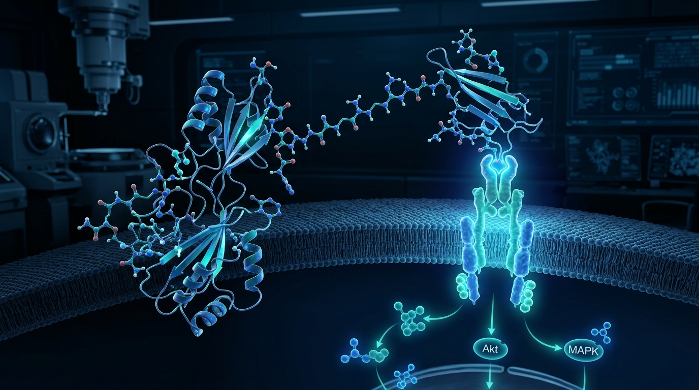
IGF-1 LR3 (Long Arg3 Insulin-like Growth Factor-1) is a synthetic analog engineered with altered binding dynamics that substantially prolongs its biological half-life, establishing it as a primary investigative tool for cellular proliferation, metabolic signaling, and myogenesis in preclinical research models.
This conclusion is supported by three primary pillars of evidence: its engineered structural resistance to inhibitory binding proteins, its heightened capacity to stimulate anabolic and mitogenic downstream pathways, and its established utility across in-vitro and animal models of tissue regeneration.
1. Engineered Structural Modifications Provide Superior Pharmacokinetics
The core utility of IGF-1 LR3 in laboratory settings stems from targeted molecular engineering designed to bypass the rapid clearance mechanisms that restrict native IGF-1.
- In-Vitro & Biochemical Evidence: Native IGF-1 is a 70-amino-acid peptide. IGF-1 LR3 contains 83 amino acids, incorporating a 13-amino-acid N-terminal peptide extension (MFPAMPLLSLFVX) and an amino acid substitution of Arginine (Arg) for Glutamic Acid (Glu) at position 3. Structural assays show this substitution significantly reduces affinity for insulin-like growth factor-binding proteins (IGFBPs)—specifically IGFBP-1 through IGFBP-6—by over 100-fold compared to native IGF-1.
- Animal Pharmacokinetic Evidence: In rodent and bovine pharmacokinetic evaluations, free native IGF-1 exhibits a serum half-life of less than 20 minutes due to rapid sequestration and renal filtration. In contrast, IGF-1 LR3 maintains circulation with an extended biological half-life estimated between 20 to 30 hours, ensuring sustained receptor engagement in experimental environments without requiring continuous infusion.
2. Potent Activation of Anabolic and Mitogenic Signaling Cascades
Because IGF-1 LR3 remains unbound in solution, it exerts continuous, potent activation of receptor-mediated intracellular pathways governing growth and cell survival.
- In-Vitro Evidence: In cultured murine myoblasts (such as C2C12 cell lines), IGF-1 LR3 binds to the IGF-1 Receptor (IGF-1R) with high specificity, triggering the phosphorylation of IRS-1 and activating the PI3K/Akt/mTOR pathway. This cascade markedly upregulates protein translation, satellite cell proliferation, and myogenin expression compared to equimolar concentrations of standard recombinant IGF-1. Additionally, activation of the MAPK/ERK pathway contributes to accelerated cellular mitosis.
- Animal Evidence: Murine studies have demonstrated that administration of IGF-1 LR3 stimulates measurable increases in muscle protein synthesis, total nitrogen retention, and organ weight, highlighting its systemic anabolic potency in vivo.
- Anecdotal Claims vs. Clinical Gaps: Outside controlled preclinical studies, anecdotal reports from unauthorized athletic circles claim significant localized muscle hypertrophy (hyperplasia) and accelerated recovery. However, there are no robust human clinical trial data evaluating the safety, efficacy, or long-term systemic impact of IGF-1 LR3 in humans, leaving these claims scientifically unvalidated.
3. Broad Preclinical Utility in Metabolic and Regenerative Models
The compound’s stability and receptor potency make it a versatile reagent for investigating tissue repair, nutrient partitioning, and cell survival under stress.
- In-Vitro Evidence: Cell culture models demonstrate that IGF-1 LR3 stimulates glucose uptake through the translocation of GLUT4 transporters to the cell membrane independently of high insulin levels. It also demonstrates anti-apoptotic properties by downregulating pro-apoptotic markers such as Bad and Caspase-3 in stressed neuronal and endothelial cell lines.
- Animal Evidence: In rodent models of gut mucosal damage, severe burn trauma, and catabolic wasting, IGF-1 LR3 administration accelerated epithelial wound closure, restored mucosal architecture, and mitigated critical weight loss.
- Hypotheses & Research Precautions: Preclinical researchers hypothesize that due to its persistent IGF-1R stimulation and anti-apoptotic signaling, prolonged exposure may elevate the risk of mitogenesis in pre-neoplastic cells or induce acute hypoglycemia via cross-reactivity with insulin receptors at elevated concentrations. These hypotheses reinforce the requirement for strict laboratory containment and dosage control in ongoing investigations.