In the field of peptide bioregulator research, Pinealon and Epithalon represent two of the most frequently examined synthetic ultrashort peptides. Both molecules emerged from the research framework pioneered by Vladimir Khavinson and colleagues at the St. Petersburg Institute of Bioregulation and Gerontology. However, commercial peptide discussions frequently blur their scientific distinctions, treating them interchangeably as generalized anti-aging compounds. In published scientific literature, Pinealon is an entirely distinct molecule with a different structural composition, primary tissue affinity, and proposed molecular mechanism than Epithalon.
Understanding the distinction between Pinealon and Epithalon requires evaluating their biochemical designs. While Epithalon is investigated primarily for systemic cellular senescence and pineal-mediated endocrine regulation, Pinealon is specifically evaluated as a neuroprotective agent targeted at the central nervous system. This comparative analysis examines the laboratory evidence, proposed epigenetic pathways, and translational limitations that separate these two bioregulators.
Key Differences: Molecular Structure and Tissue Origins
At the chemical level, Pinealon and Epithalon belong to distinct structural peptide classes. Pinealon is a synthetic tripeptide consisting of three amino acid residues: L-glutamic acid, L-aspartic acid, and L-arginine (abbreviated in scientific literature as Glu-Asp-Arg or EDR). It possesses a molecular formula of C15H26N6O8 and a low molecular weight of approximately 418.4 Da. This ultra-compact profile gives Pinealon high aqueous solubility and enables it to pass readily across biological barriers, including the blood-brain barrier and the nuclear envelope.
In contrast, Epithalon (also referred to as Epitalon or AEDG) is a synthetic tetrapeptide consisting of four amino acids: L-alanine, L-glutamic acid, L-aspartic acid, and glycine (Ala-Glu-Asp-Gly). While both molecules share a central acidic core of glutamic acid and aspartic acid (Glu-Asp), their flanking residues confer different conformational dynamics and binding behaviors.
A widespread misconception involves the physiological origins of their parent extracts:
- Pinealon (EDR): Despite an ambiguous name that suggests a pineal origin, research reviews indicate that the EDR sequence was identified as an active bioregulatory motif derived from Cortexin, an animal polypeptide extract harvested from cerebral cortex tissue. Consequently, its preclinical investigation has focused heavily on cortical and hippocampal neurons.
- Epithalon (AEDG): Epithalon was designed to replicate the primary biological activity of Epithalamin, a peptide complex extracted directly from bovine pineal gland tissue, with initial studies centering on pineal restoration and neuroendocrine rhythms.
Mechanism of Action: Pinealon in Neurons vs. Epithalon in Cellular Aging
The molecular targets identified for Pinealon diverge substantially from those characterized for Epithalon, reflecting their different experimental pathways.
Pinealon: Neuronal Gene Regulation and Oxidative Resilience
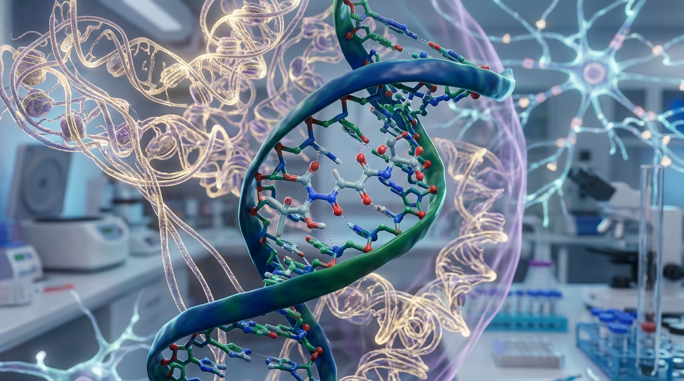
Research into Pinealon emphasizes direct cytoprotection in neural tissue. Laboratory biophysical models propose that ultrashort peptides such as EDR enter cells via passive diffusion or peptide transporters and interact directly with nuclear material. Studies using fluorescent labeling and nuclear magnetic resonance (NMR) spectroscopy indicate that EDR can bind into the major and minor grooves of double-stranded DNA, specifically engaging core promoter sequences of genes involved in neuronal differentiation, neurotransmission, and oxidative defense.
Preclinical in vitro assays demonstrate that Pinealon modulates the expression of endogenous antioxidant enzymes, including superoxide dismutase (SOD1 and SOD2), thereby suppressing the intracellular accumulation of reactive oxygen species (ROS). Furthermore, experimental models of neurodegeneration suggest Pinealon downregulates pro-apoptotic signaling—such as caspase-3 activation—while maintaining structural cytoskeletal proteins in cortical and cerebellar neurons.
Epithalon: Telomerase Activation and Endocrine Modulation
Epithalon's primary mechanistic footprint centers on the regulation of telomere dynamics and neuroendocrine homeostasis. Early publications reported that AEDG could induce the expression of human telomerase reverse transcriptase (hTERT) and promote telomerase enzymatic activity in somatic cell cultures, potentially overcoming the replicative Hayflick limit in senescent cells. Additionally, Epithalon acts within the pineal gland to stimulate the synthesis of melatonin, theoretically re-entraining degraded circadian oscillations and normalizing hypothalamic-pituitary-adrenal (HPA) axis balance in aging animal models.
Preclinical and Clinical Evidence: What the Studies Show
Neither compound has achieved authorization from major regulatory agencies, but their respective preclinical dossiers address different experimental questions.
Pinealon Evidence: Synaptic Preservation and Brain Stress Models
Experimental studies evaluating Pinealon have established several noteworthy neurobiological endpoints:
- Dendritic Spine Protection: In transgenic mouse models of Alzheimer's disease (such as 5xFAD models), administration of Pinealon was found to preserve mushroom-shaped dendritic spines in hippocampal neurons. Loss of these mature synaptic structures correlates directly with memory impairment, indicating that EDR may sustain synaptic architecture under beta-amyloid stress.
- Human-Derived Induced Neurons: A 2024 study published in the International Journal of Molecular Sciences evaluated EDR in induced neurons transdifferentiated from elderly human dermal fibroblasts. The study observed that EDR reduced oxidative DNA damage and stimulated primary dendrite branching, providing evidence of cellular response in human-derived neural phenotypes.
- Hypoxic and Prenatal Insults: In rodent models of maternal hyperhomocysteinemia, prenatal administration of Pinealon protected the developing fetal brain from oxidative injury, improving subsequent spatial learning parameters in the offspring.
- Human Observational Data: Small, open-label Russian clinical reports evaluated oral Pinealon in patients with traumatic brain injury sequelae or cerebrovascular insufficiency. While authors reported subjective improvements in cognitive performance and normalization of electroencephalographic (EEG) alpha rhythms, these studies were not randomized, double-blind, or placebo-controlled by modern International Council for Harmonisation (ICH) standards.
Epithalon Evidence: Senescence and Longevity Endpoints
Epithalon's primary evidence base consists of longevity cohorts conducted across rodents and non-human primates. In long-term rodent experiments, AEDG administration was associated with a reduction in spontaneous mammary and leukemia-like tumor incidence and modest extensions in median lifespan. In aged primates (Macaca mulatta), Epithalon restored the diurnal rhythm of melatonin and evening cortisol production toward profiles seen in younger animals. However, human evidence remains limited to preliminary non-blinded trials measuring immune parameters and cardiovascular markers.
Methodological Limitations and the Single-Network Problem
When analyzing Pinealon—and comparing it to Epithalon—independent evaluators emphasize notable methodological vulnerabilities across the scientific literature:
- Research Network Concentration: Virtually the entire primary literature on Pinealon and Epithalon originates from a single research network based at the St. Petersburg Institute of Bioregulation and Gerontology. Robust independent replication in international, peer-reviewed laboratories remains scarce.
- Direct Comparative Trials: No head-to-head clinical trials directly compare Pinealon and Epithalon under controlled therapeutic conditions. Published contrasts rely primarily on cross-study comparisons between independent datasets.
- Surrogate Endpoints: Much of the mechanistic data for Pinealon relies on computer docking models, in vitro radical suppression, or surrogate histological markers (such as spine counts or dendritic length) rather than functional reversal of neurological disease in humans.
- Inconsistent Formulations: Literature descriptions alternate between oral preparations, intramuscular injections, and sublingual delivery without providing validated pharmacokinetic or bioavailability profiles.
Regulatory Status and Research Availability
Neither Pinealon nor Epithalon is approved by the U.S. Food and Drug Administration (FDA), the European Medicines Agency (EMA), or Health Canada for the prevention, diagnosis, or treatment of any medical condition. In Western jurisdictions, both substances are classified exclusively as investigational research biochemicals intended for laboratory experimentation and are not approved for human clinical use or dietary supplementation.
Frequently Asked Questions
What is the primary functional difference between Pinealon and Epithalon?
Pinealon is a tripeptide (Glu-Asp-Arg) focused on neuroprotection, neuronal survival, dendritic spine morphology, and brain oxidative stress. Epithalon is a tetrapeptide (Ala-Glu-Asp-Gly) investigated primarily for telomerase activation, systemic cellular senescence, and pineal melatonin synthesis.
Why is Pinealon associated with the brain if its name suggests the pineal gland?
Although the name suggests a pineal connection, scientific literature indicates that Pinealon was designed based on peptide fractions identified in Cortexin, an extract of cerebral cortex tissue. Most of its published effects specifically involve cortical and hippocampal neurons rather than pinealocyte biology.
Are Pinealon and Epithalon approved as pharmaceuticals?
No. Neither peptide has received FDA approval or marketing authorization in North America or Western Europe. They remain unapproved investigational compounds restricted to in vitro and animal laboratory research.
Can Pinealon findings in rodents be generalized to human clinical medicine?
No. While rodent and cell culture studies show positive effects on oxidative stress and dendritic preservation, rodent models do not duplicate the full complexity of human neurodegenerative diseases. Controlled, double-blind clinical trials are necessary before determining human efficacy or safety.
Research Summary
Pinealon (Glu-Asp-Arg) and Epithalon (Ala-Glu-Asp-Gly) represent distinct branches of peptide bioregulator research. While Epithalon is oriented toward systemic longevity, telomere maintenance, and circadian endocrine rhythms, Pinealon functions in experimental models as a targeted neuroprotective agent that supports dendritic spine stability, modulates antioxidant gene pathways, and protects against neuronal oxidative stress. The evidence for both compounds remains predominantly preclinical, originating largely from a single academic institution without independent phase III clinical trials. Neither agent holds regulatory approval for therapeutic use.
References
- Khavinson, V., Linkova, N., Kozhevnikova, E., & Trofimova, S. (2020). EDR Peptide: Possible Mechanism of Gene Expression and Protein Synthesis Regulation Involved in the Pathogenesis of Alzheimer's Disease. Molecules, 26(1), 159. https://pubmed.ncbi.nlm.nih.gov/33396470/
- Kraskovskaya, N., Linkova, N., Sakhenberg, E., et al. (2024). Short Peptides Protect Fibroblast-Derived Induced Neurons from Age-Related Changes. International Journal of Molecular Sciences, 25(21), 11363. https://pubmed.ncbi.nlm.nih.gov/39518916/
- Khavinson, V., Ribakova, Y., Kulebiakin, K., et al. (2011). Pinealon increases cell viability by suppression of free radical levels and activating proliferative processes. Rejuvenation Research, 14(5), 535–541. https://pubmed.ncbi.nlm.nih.gov/21978084/
- Arutjunyan, A., Kozina, L., Stvolinskiy, S., et al. (2012). Pinealon protects the rat offspring from prenatal hyperhomocysteinemia. International Journal of Clinical and Experimental Medicine, 5(2), 179–185. https://www.ncbi.nlm.nih.gov/pmc/articles/PMC3342713/
- Fedoreyeva, L. I., Kireev, I. I., Khavinson, V. K., & Vanyushin, B. F. (2011). Penetration of short fluorescence-labeled peptides into the nucleus in HeLa cells and in vitro specific interaction of the peptides with deoxyribooligonucleotides and DNA. Biochemistry (Moscow), 76(11), 1210–1219. https://pubmed.ncbi.nlm.nih.gov/22117547/